Number of Bones in Human Body Skeleton Facts DK Find Out
The tibia is also known as the shinbone, and is the second largest bone in the body. There are two bones in the shin area: the tibia and fibula, or calf bone. The fibula is smaller and thinner.
What are Bones Made of? Bones in the Human Body Wiki
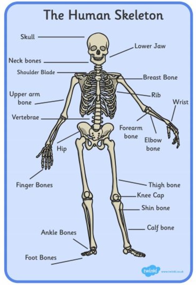
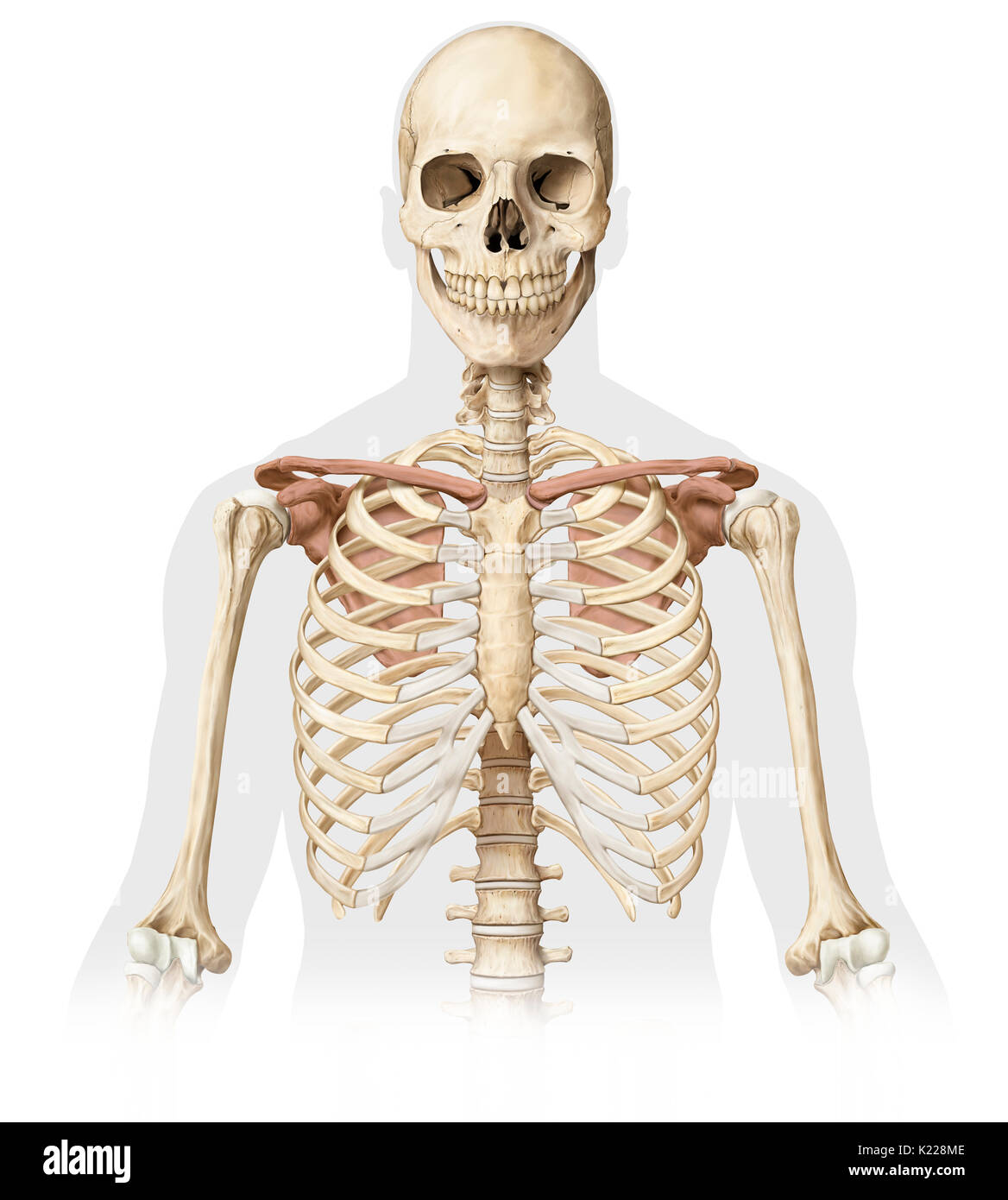
The clavicle, commonly known as the collarbone, is a slender, S-shaped, modified long bone located at the base of the neck. It is the only long bone of the body that lies horizontally. The term clavicle comes from the Latin word ' clavicula ', meaning 'little key', as its shape resembles an old-fashioned key.
File603 Anatomy of Long Bone.jpg Wikimedia Commons
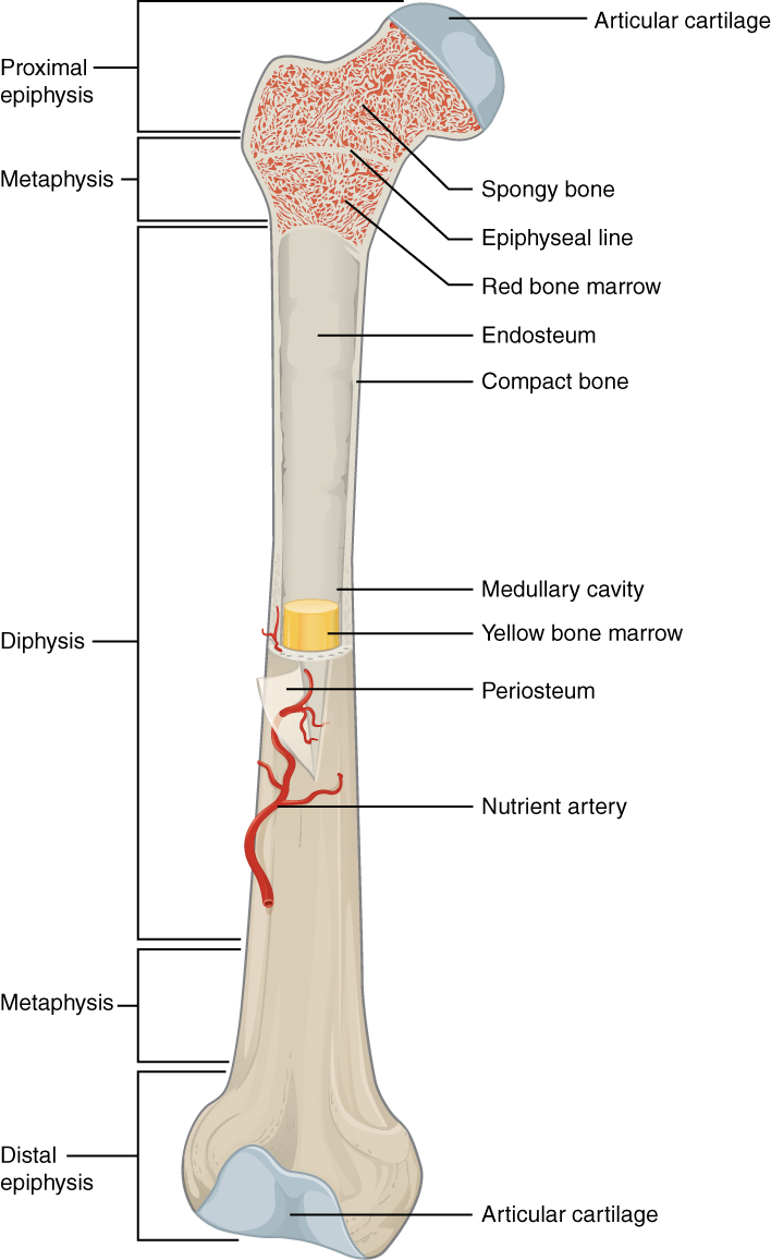
The skeletal system includes all of the bones and joints in the body. Each bone is a complex living organ that is made up of many cells, protein fibers, and minerals. The skeleton acts as a scaffold by providing support and protection for the soft tissues that make up the rest of the body. The skeletal system also provides attachment points for.
3D model Bone Marrow Anatomy TurboSquid 1739292
The vertebral column, commonly known as the spine, spinal column, or backbone, is a flexible hollow structure through which the spinal cord runs. It comprises 33 small bones called vertebrae, which remain separated by cartilaginous intervertebral discs. The vertebral column forms the axial skeleton, skull bones, ribs, and sternum.
Biology Reading Flashcards Bones Ask A Biologist
The hip joint is a ball-and-socket synovial joint formed between the os coxa (hip bone) and the femur. A round, cup-shaped structure on the os coxa, known as the acetabulum, forms the socket for the hip joint. The rounded head of the femurmycontentbreak forms the ball of the joint. Hyaline cartilage lines both the acetabulum and the head of the.
Upper body bones hires stock photography and images Alamy
The femur is the thigh bone and is the largest bone in the human body. A fractured femur can be life-threatening due to blood loss. Learn more about its anatomy, function, and treatment options from Verywell Health, a trusted source of health information. You can also explore other topics related to the skeletal, nervous, and respiratory systems, as well as common skin and liver conditions.
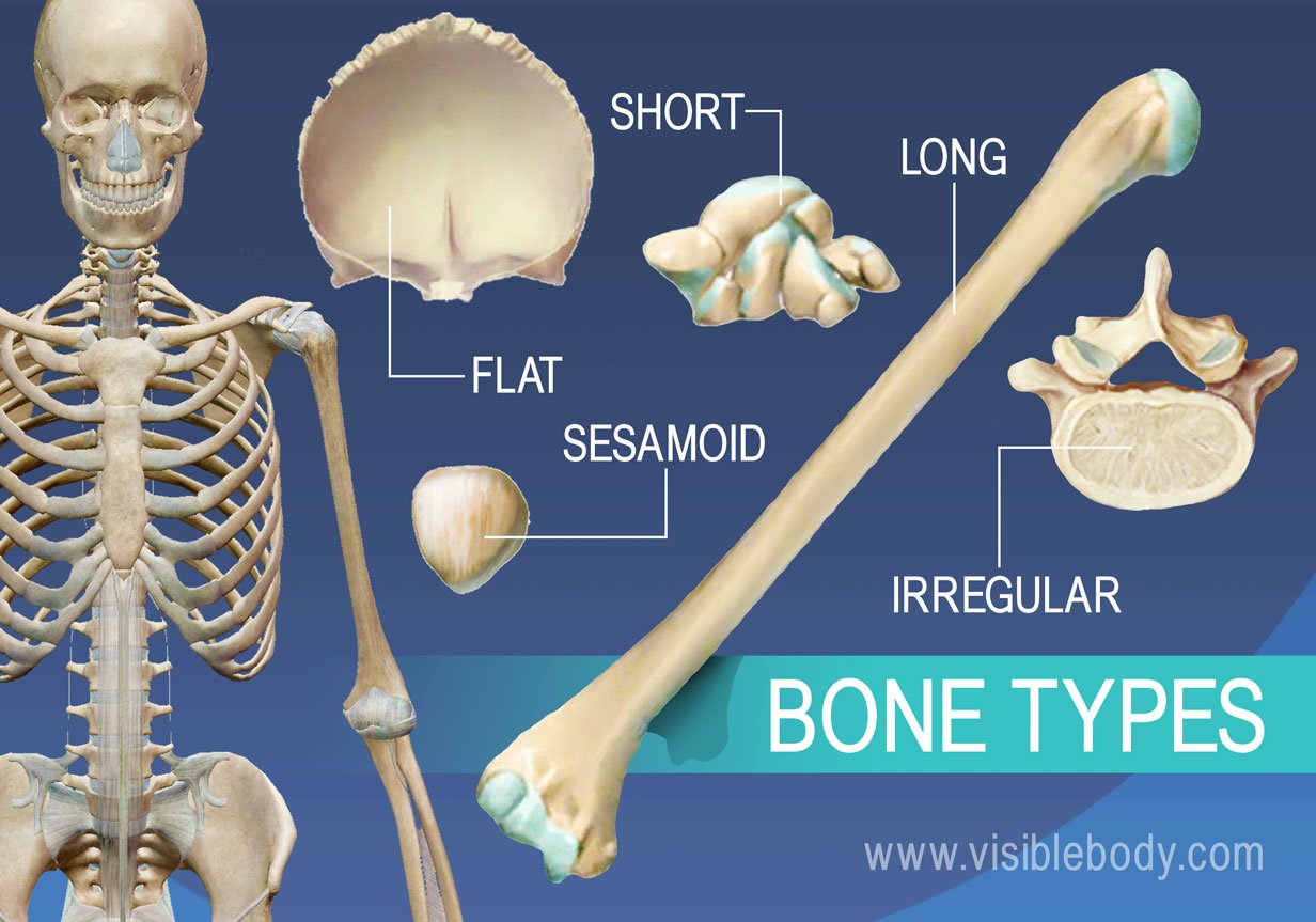
Types of Bone Biology for Majors II

The sternum, or breastbone, is a flat bone at the front center of the chest. The ribs and sternum make up what is called the 'ribcage.'. The ribcage protects the lungs, blood vessels, and.
Show Me the Bone Reconstructing Prehistoric Monsters in Britain and America
Summary. The skeletal system is made up of your bones, ligaments, and cartilage. Though its main function is to provide structural support for the body, it also stores important minerals—such as calcium—forms red blood cells, and protects your internal organs. The skeletal system can break down into two main categories—the axial skeleton.
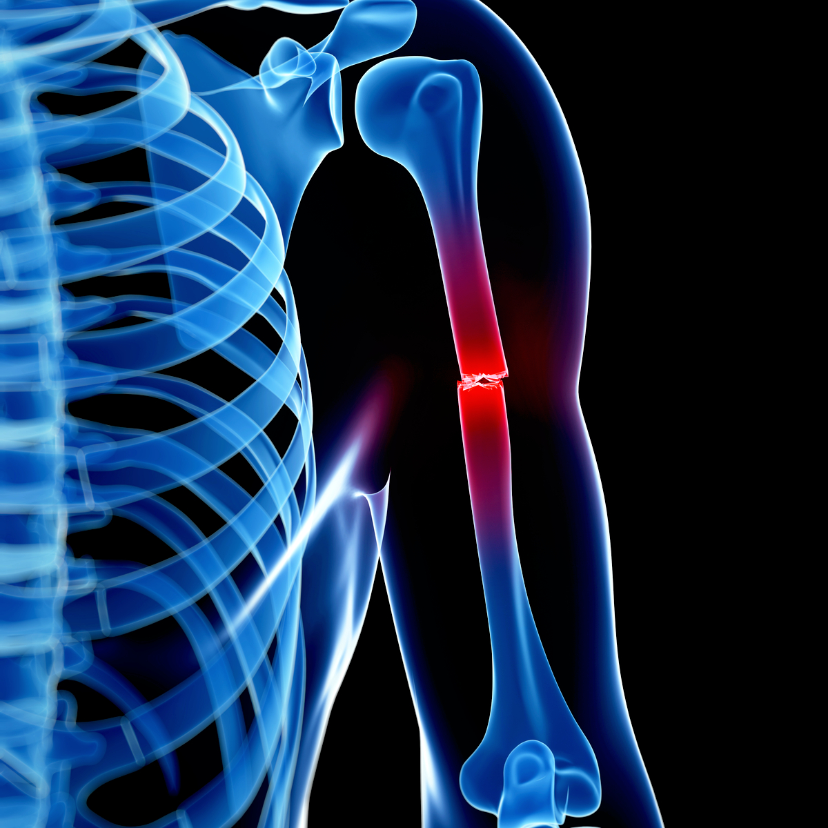
Why was a fracture missed on an x ray? The Medical Consultant
Human skeleton, muscles and blood vessels, illustration. Browse Getty Images' premium collection of high-quality, authentic Human Skeleton Anatomy stock photos, royalty-free images, and pictures. Human Skeleton Anatomy stock photos are available in a variety of sizes and formats to fit your needs.
Diagram of human bone anatomy Stock Vector Image by ©roxanabalint 18985777

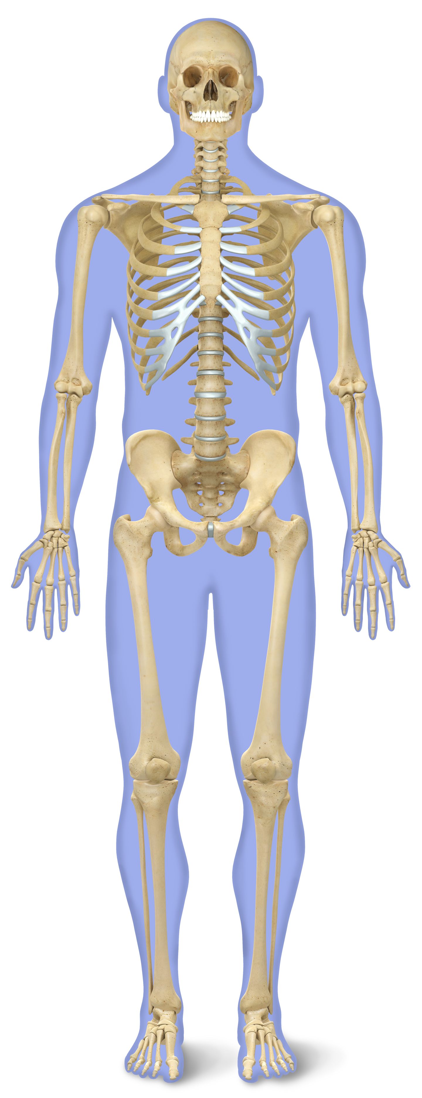
The human skeleton of an adult usually consists of around 206 bones, depending on the counting of sternum (which may alternatively be included as the manubrium, body of sternum, and the xiphoid process). It is composed of 270 bones at the time of birth, but later decreases to 206: 80 bones in the axial skeleton and 126 bones in the appendicular skeleton. 172 of 206 bones are part of a pair and.
Bone Anatomy Ask A Biologist

The hip joint is a ball-and-socket type joint and is formed where the thigh bone (femur) meets the pelvis. The femur has a ball-shaped head on its end that fits into a socket formed in the pelvis, called the acetabulum. Large ligaments, tendons, and muscles around the hip joint hold the bones (ball and socket) in place and keep it from dislocating.
Bones in the Hip JOI Jacksonville Orthopaedic Institute

Medical school student and professor discuss human spine model. of 100. Browse Getty Images' premium collection of high-quality, authentic Human Bones Anatomy stock photos, royalty-free images, and pictures. Human Bones Anatomy stock photos are available in a variety of sizes and formats to fit your needs.
Overview of Skeleton Learn Skeleton Anatomy
According to the American College of Rheumatology, 70% of people over 70 have X-ray evidence of osteoarthritis . X-ray pictures of osteoarthritis can show joint cartilage deterioration, compressed spinal discs, bony nodules, and other evidence of joint damage. This article discusses osteoarthritis symptoms, diagnosis, and treatment.
Human Bone Anatomy Labeled City Distributers Human Bones Resource site for teachers and

The sternum, commonly known as the breastbone, is a long, narrow flat bone that serves as the keystone of the rib cage and stabilizes the thoracic skeleton. Several muscles that move the arms, head, and neck have their origins on the sternum. It also protects several vital organs of the chest, such as the heart, aorta, vena cava, and thymus.
Home Anatomy & Physiology LibGuides at COM Library

Femur. The femur is the only bone located within the human thigh. It is both the longest and the strongest bone in the human body, extending from the hip to the knee. Important features of this.
Bone Anatomy and Function

Crossed Bones on a White Background. of 100. United States. Browse Getty Images' premium collection of high-quality, authentic Human Bone stock photos, royalty-free images, and pictures. Human Bone stock photos are available in a variety of sizes and formats to fit your needs.
.